Tag Archives: Winkler Center

3D Imaging Workshop in the Lab

This past December, the Lab hosted a small 3D Imaging workshop for a group of digital imaging colleagues. The workshop was led by UC School of Art professors, Jordan Tate and John-David Richardson. Colleagues from Ohio University Libraries, the Ohio State University Libraries, Library of Michigan, Veterans Affairs History Office in Dayton and UC Libraries were able to join us for this amazing opportunity to learn practical, high-resolution 3D imaging techniques using a simple setup.

Person kneeling behind a camera with four other people standing behind him.
Jordan demonstrating to the group how to set up a low-angle shot.

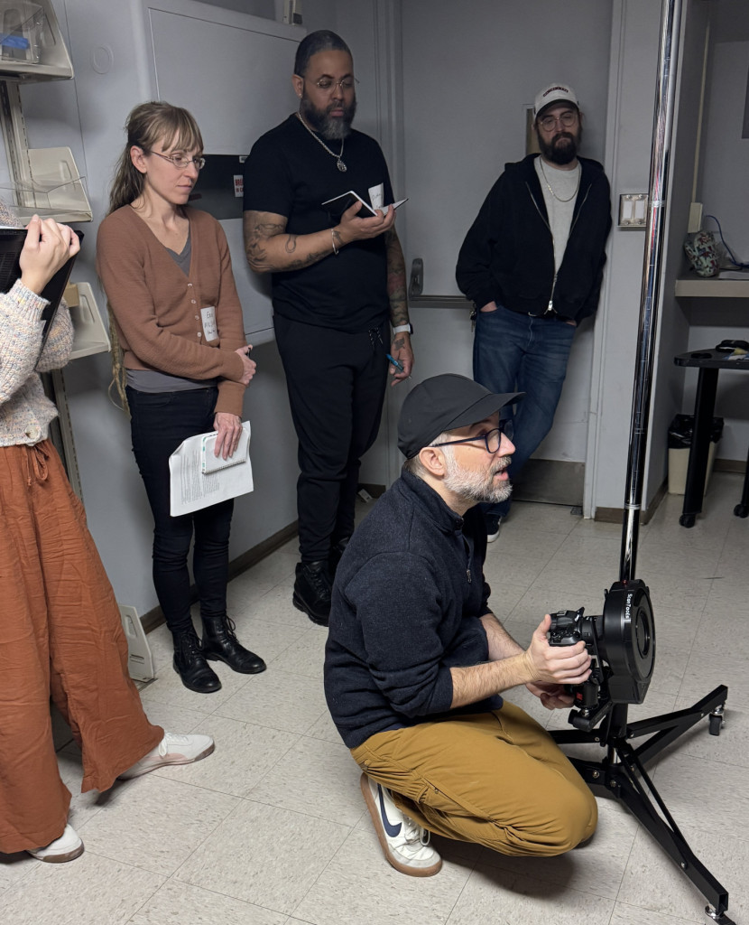
Prior to the workshop, I had worked with UC Libraries special collections curators to attain a selection of various objects for imaging ranging from cuneiform tablets to plaster busts. For the workshop, we began with a painted wooden Nigerian statue from the Winkler Center. We walked through the imaging process first, using a fairly standard DSLR manual setup and a ring flash with a polarized filter. The object was placed on a motorized turn-table which allows you to determine how many rotations or shots you want per angle.

Yoruba statue, from the Winkler Center, being photographed on the motorized turntable.

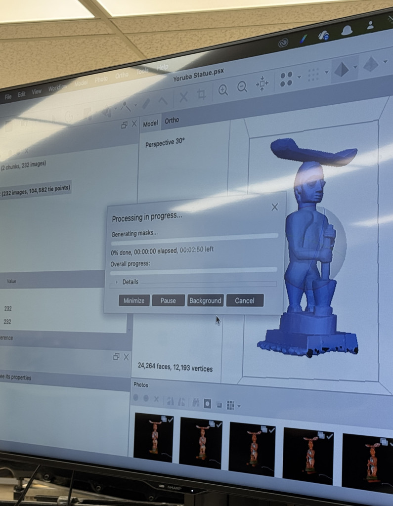
Once all the images were captured, we moved onto the processing portion, using Agisoft Metashape Professional Educational edition.

Screen with 3D imaging software showing a model of an African statue being processed.
The model of the Yoruba statue being processed in Agisoft Metashape software.

After lunch we ran through another imaging and processing session, to reinforce what we had learned in the morning. Overall the day was incredibly successful and I am beyond grateful to Jordan and John-David for sharing their time, knowledge and passion for 3D imaging with the group. They truly made the imaging capture and processing experience fun and attainable!

Following the workshop, fellow Assistant Conservator, Catarina, and I were able to get some 3D imaging practice in; using what we had learned in the workshop and putting it into action. This was made possible by two very important factors: first, Jordan had graciously loaned us his equipment to use before the ’26 Spring semester began, including his polarized flash ring, and second, we had a couple of slower days, following our special collections returns and prior to winter break, which afforded both Catarina and I this time to learn and hone this new technique.

The combat medic statue from the Winkler Center being photographed.

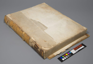
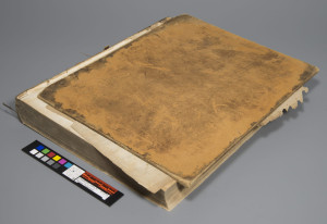
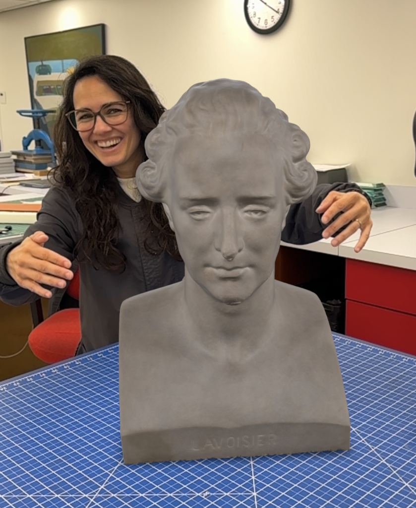
In the end, Catarina and I ended up imaging three additional objects, including a Gothic manuscript from the Archives and Rare Books Library, a plaster bust of Antoine Lavoisier from the Oesper Collection, and a combat medic statue from the Winker Center.

Again, thanks to Jordan’s immense generosity, he has hosted the 3D models of the objects we captured on his site, so that they can be shared more broadly with a wider audience. If you can click on the links below (whether with computer, phone or tablet) you can view and manipulate the 3D models of each object, as well as read more about the object:

Again, a huge thank you to Jordan and John-David for their eagerness to share their knowledge during the workshop, and to Jordan for his ongoing generosity and collegiality. And, as always, thank you to our curators for supplying us with an array of objects for the workshop, and to our department head, Holly Prochaska, for her perennial support and advocacy.

Woman attempting to hug a 3D model of Antoine Lavoisier (bust).
Catarina attempting to hug the Lavoisier bust model. This model is being projected from an iPhone, allowing it to be “placed” on the table.

Jessica Ebert – Assistant Conservator

Illustrated Human Opening Reception – 11/16

Please join us for the second lecture in the six-part series – “Making the Fabrica: The Illustrations, Printing, Binding & Publication.” Award-winning cultural historian Dániel Margócsy, PhD, University of Cambridge, will describe the creation of the “Fabrica.” Dr. Margócsy will be joined by Gabrielle Fox, a Cincinnati book binding and preservation expert, who will discuss the bindings of the first and second editions that will be on public display in the Donald C. Harrison Health Sciences Library.

The lecture, free and open to the public, will be held Tuesday, Nov. 16 at 5:30 p.m. in Kresge Auditorium, 231 Albert Sabin Way. In-person activities will be provided under CDC guidelines or local COVID-19 restrictions, with the well-being of all guests remaining the top priority. View UC’s current COVID-19 updates.  For those not wishing to attend in person, the lectures will be live streamed via Zoom.

Register to attend the lecture.

Off the shelf and into the lab – additional resources

Poster of the Cecil Striker Lecture Series featuring Preservation Lab staff in action.

Thanks to everyone who attended the May 6th Striker Lecture, Off the Shelf and into the Lab: Medical History, Preservation, & the University of Cincinnati Libraries’ Adopt-A-Book Program.

If you missed the program, a recording is now available.

Additionally, you might want to check out a couple additional blogs that we mentioned in the question and answer session, such as our favorite treatments post and rehousing of the Winkler Center’s prosthetic ear post. Also, to compliment the program, we created a video tour of the lab available on our YouTube Channel.

Screen shot of the video Virtual Tour of the Preservation Lab.
Check out the Preservation Lab’s YouTube channel – https://www.youtube.com/channel/UCeTBLuvqGe0SouIy2bgiUnw

Holly Prochaska (UCL) — Preservation Librarian

May 6th Webinar – Off the Shelf and into the Lab

Join Holly and Ashleigh this Thursday at 7pm (EST) for the 3rd lecture in the Cecil Striker Webinar series, Off the Shelf and into the Lab: Medical History, Preservation, & the University of Cincinnati Libraries’ Adopt-A-Book Program.

Ashleigh and Holly will talk about the work we do in the Lab and UC Libraries’ Adopt-A-Book program.

Zoom details:
https://ucincinnati.zoom.us/j/93403337138

Meeting ID: 934 0333 7138

More details about the Adopt-A-Book Program and items up for adoption can be found at: https://libapps.libraries.uc.edu/adoptabook/

Jessica Ebert (UCL) – Conservation Tech & Photographic Documentation Specialist

A Monster of a Treatment

When this mid-19th century Treatise on Operative Surgery came to The Preservation Lab, little did we imagine the twists and turns the treatment would take. We certainly did not expect the treatment to turn into a Frankenstein’s monster, pulling inspiration from three different types of binding structures, although this monster found more successful results than Victor Frankenstein’s.

This 1844 full leather tight-back binding came to us from The Henry R. Winkler Center. The binding was in disarray with both the front and back boards having become fully detached, with only the back board remaining. Very few spine fragments remained, but there was clear evidence of false raised bands. The leather that remained on the spine and back board was severely red rotted. The textblock was sewn on recessed cords and remained beautifully intact. The textblock consists of 360 pages containing descriptions of surgical procedures, as well as 80 illustrated plates depicting some of these techniques.
Prior to any rebinding, the placement of the false raised bands was measured and recorded; the book’s spine was cleaned; the front and back flyleaves were mended and hinged; the back cover was housed in a polyester four-flap; new endsheets were sewn onto the textblock; and the spine was lined. Several of the linings attached will bring us to the first bookbinding structure that inspiration was pulled from…

The Split Board Binding: Because of the large size of the book, a split board binding attachment was chosen to add strength to the board attachment. After the spine of the textblock was lined with Japanese tissue and wheat starch paste, creating a reversible layer, a secondary spine lining with flanges was created out of linen and attached with Jade 403 PVA. This linen is an integral part of the split board structure, as it, along with the first endsheet and sewing supports, will create the “laminated flange” that will be pasted in between the two boards (hence, split board). This flange and split board structure can be seen in the uppermost book model in the stack pictured above.
Now that the book has the structure and stability of a split board binding, we can pull from another binding structure to attach the raised supports…

The “Baggy Back” Binding: A “baggy back” structure, also referred to as a conservation case, is what will allow the spine of the binding to be formed to the spine of the textblock, without being adhered directly to it. Another way to think about it is an alternative to a hollow tube structure. Sized linen was non-adhesively shaped over the spine of the textblock, and was pasted in between the split boards on top of the laminated flanges. The open book model in the image on the left shows what a typical “baggy back” structure looks prior to casing the textblock. Comparing that to how the linen can be integrated into the split board structure, we start to see our Frankenstein’s monster take shape.
To prep for the final stages of rebinding, some elements were added to stay true to the original structure of the book. False raised bands were cut and shaped out of 4-ply mat board to mimic the shape and location of the original false raised bands. Hollow tube segments were also attached in between the false raised bands.

These spine elements will finally be covered up using inspiration from one final book structure…
The Quarter Linen Binding: The book was decided to be finished as a quarter linen binding, instead of a more historical full leather or quarter leather binding for conservation purposes. The third model referenced, shown above, depicts the end result of this style of binding. In this structure the linen is stretched over the spine of the book to add the definition of the raised bands, or in the case of this treatment, false raised bands. The linen also allows for a strong attachment between the textblock and the boards. The hollow tubes laid between the bands allow for a better opening once the linen is in place.
During this treatment the linen was stretched over the spine of the book and onto the front and back boards, making sure the linen is securely and evenly applied around all of the spine elements. The book was then secured in a tying press to ensure definition of the bands.

Turn-ins were properly secured, and the linen was trimmed to the proper length on the boards. A brown book cloth was used to cover the remainder of the boards, the color chosen to mimic the color of the original leather used in the binding. The internal flanges from the split board binding structure (as seen in the right image above), along with the pastedowns, were applied to the inner boards. And no book is complete without a stamped title and a matching cloth covered clamshell.

Using three different book structures to plan this treatment does seem like a Frankenstein’s monster of a project, but the success of the treatment pays homage to the integration of bookbinding techniques we find in book structures throughout history.  And this book will certainly look like no monster when it is on display for tours at The Winkler Center.

Kasie Janssen (PLCH) – Senior Conservation Technician
Photographic Documentation by Kasie Janssen & Jessica Ebert

Traveling Objects on Loan for Exhibition


This week we had the pleasure of assessing the condition of some unusual objects not often found in a book and paper conservation lab.  The objects, owned by the Winkler Center for the History of Health Professions at UCL, consisted of a child’s leg brace, a box of polio specimen slides from the 1960’s, and a group of honorary medals.

Continue reading

An "earie" object to have in the Lab…

Recently we received an unusual item from the University of Cincinnati’s Winkler Center for the History of the Health Professions:  a prosthetic ear dating to the mid 1950’s accompanied by a small photograph and newspaper clipping depicting the patient modeling the false ear.  Fortunately, or unfortunately depending on your point of view, the ear we received in the lab was a primary model used to construct the actual prosthetic, so it would not have been worn regularly by the patient.  I have to admit this is one of the more gruesome items I’ve come across in a conservation lab.  Not because it’s a prosthetic ear, but more so because improper housing and storage conditions led to deterioration which gave the ear a very bumpy almost wart-like surface appearance…and it looks so real…

The ear and its original housing materials.  The photograph and clipping were stored in the yellow envelope.

The ear and its original housing materials. The photograph and clipping were stored in the yellow envelope.


Continue reading