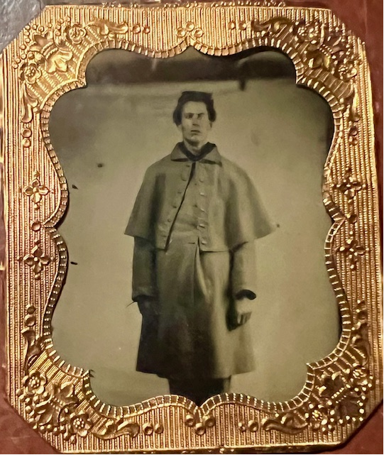
Benjamin L. Askue, Jr. during the American Civil War, circa 1860s
Frontline accounts of military conflicts provide a glimpse into the world of the war. The historical record reflects numerous descriptions of soldier’s and military doctor’s accounts of the bloodiest war ever engaged on American soil – the Civil War. The Henry R. Winkler Center for the History of the Health Professions announces the launch of the narratives of the 23rd Regiment Ohio Volunteers Infantry doctor, the Benjamin L. Askue, Jr. Civil War LettersBenjamin L. Askue, Jr. Civil War Letters on JSTOR.
Askue was born in November 1833 to Benjamin and Rowena Cordelia Askue in Ashtabula, Ohio. In 1853, he married his cousin Flavia Pritchard. The letters he wrote to Flavia during the American Civil War demonstrate that they had a happy marriage. The couple had five children together.
During the 19th century and early 20th century, physicians often received their training through apprenticeships. Askue followed this path becoming a homeopathic doctor. In 1861 he joined the 23rd Regiment Ohio Volunteers Infantry, Company B in the Union Army. Askue served as a cook, nurse, hospital steward, and in the 23rd Regiment’s infantry. He left the Union Army In July 1865. Askue returned to Ashtabula to farm and practice homeopathic medicine. He died in 1906.
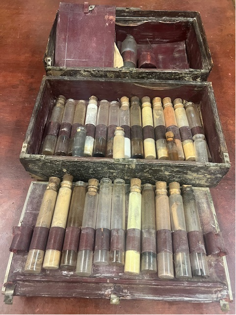
Askue’s archives and artifacts were donated to the Winkler Center. While his archives hold numerous documents and artifacts, the highlight of the collection consists of letters written to Flavia beginning in June 1861 and concluding in July 1865. He described the 23rd Regiment’s travels, battles, camp life, politics, family in Ashtabula, Ohio and Askue engaged in philosophical analysis of the era.
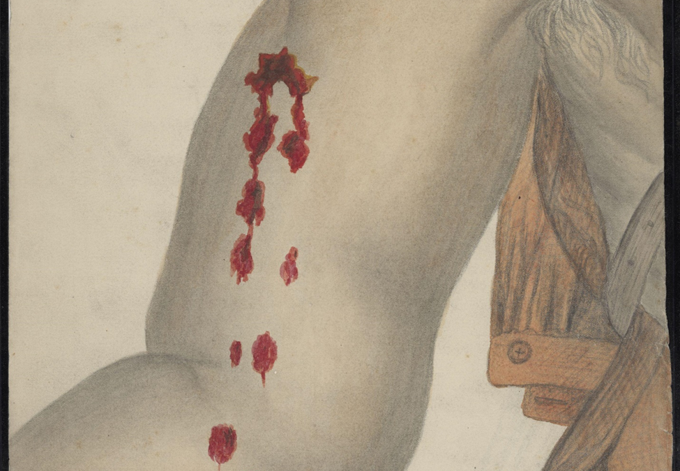
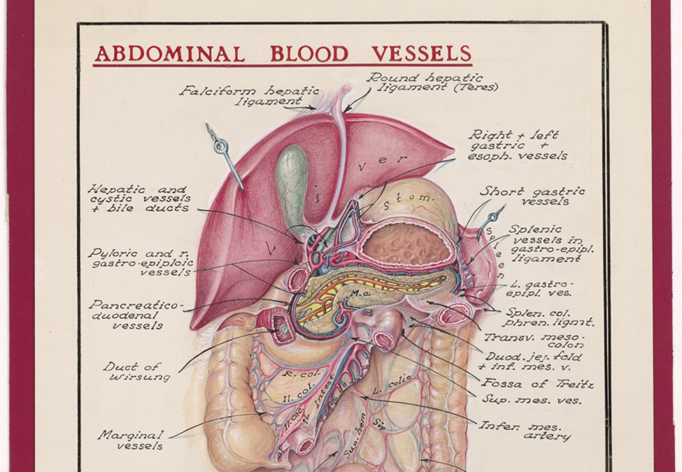
Medical illustrations and drawings are a reflection of the state of medical practice at a specific moment in time providing a visual record of science, technology, and anatomical knowledge.
The artwork of Daniel S. Young highlights the artistic and medical contributions of an American Civil War era medical illustrator in a military context. Daniel S. Young: American Civil War Medical Illustrations on JSTOR. His artistry paints a portrayal of how medical illustration informed medical professionals during the 19th century. Young’s Civil War medical illustrations were crucial in educating doctors on surgical procedures and about previously unseen wounds. While medical illustrations such as Dr. Daniel Young’s served to educate doctors they were also important in aiding veterans in their pension claims and showing how the war impacted the soldiers’ health.
Cuts along the upper arm and elbow. Stone’s River, Tennessee.Continue reading →
Founder of the University of Cincinnati, College of Medicine, School of Medical Illustration (1930-1972)
By Devhra BennettJones
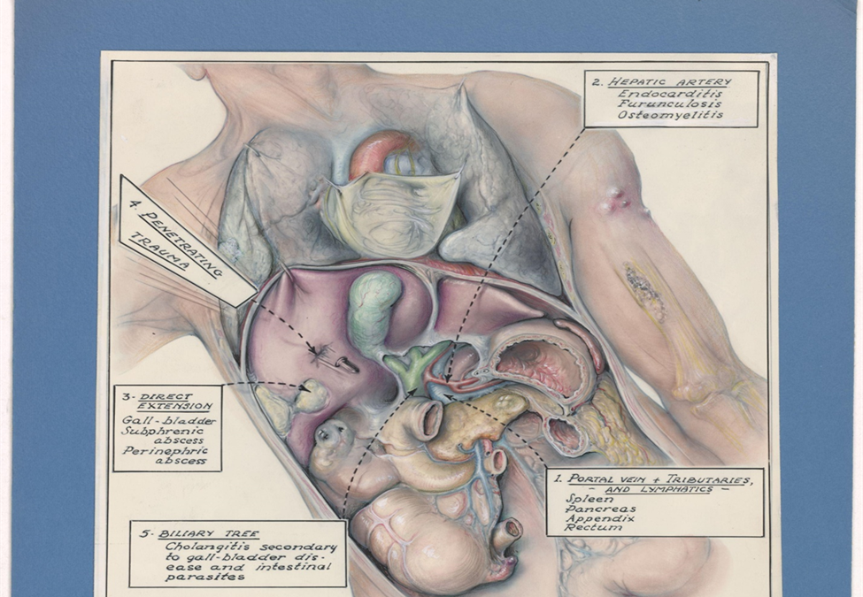
Penetrating trauma surgery
Abdominal Blood Vessels
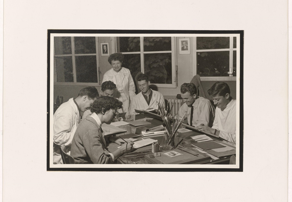
The Henry R. Winkler Center for the History of the Health Professions is pleased to announce online access to the archives of esteemed medical illustrator, professor and artist, Mary Maciel. Mary Maciel: Visionary in Medical Illustration on JSTOR. Maciel’s career at the University of Cincinnati College of Medicine, began in 1930 in the Department of Surgery and continued through 1972 when she retired from the School of Medical Illustration. She trained with the world renowned “father” of medical illustration, Max Broedel at Johns Hopkins University. By 1947 the University of Cincinnati Board of Directors authorized a new course in medical illustration taught by 25-year-old Mary Maciel.
Mary Maciel Instructing Medical Illustration Students
Her artistic talent and academic administrative skills led the field world-wide. She set the standard that applicants to the UC School of Medical Illustration must possess at least four, and preferably five years of course work in general art and training in science. She only allowed the admission of a maximum of two students annually. In 1948 Maciel organized a school of medical illustration in Portugal. In 1951 she established a school of medical illustration at the University of Lyons, France. By the 1950s the University of Cincinnati School of Medical Illustration was one of four North American universities with programs in medical illustration along with Johns Hopkins University, the University of Georgia, and the University of Toronto.
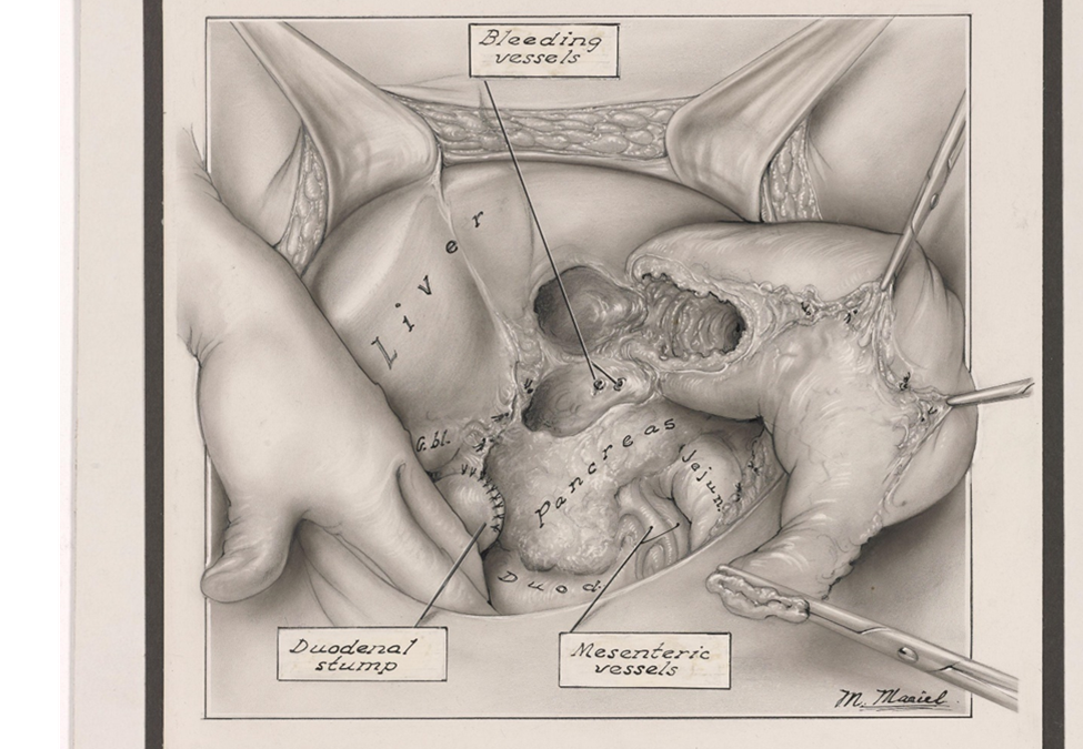
Liver Surgery
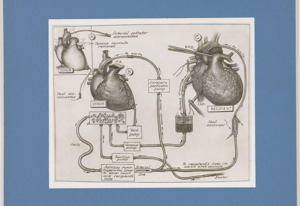
Maciel’s expertise was recognized around the globe. She often spent the summer months abroad working in the field. In 1957 she taught students of medical illustration in Finland and was awarded a four-month Fulbright professorship at the University of Strasbourg. In 1958 Mary Maciel presented lectures and medical illustration demonstrations at the University of Helsinki. She served as a consultant at medical centers in Denmark, Sweden and Norway. In 1963 Maciel visited medical schools in Brazil, Uruguay, and Argentina. She was a visiting professor at the University of Rio de Janeiro and the University of Buenos Aires. In 1968 Maciel was invited to work with the esteemed Dr. Christiaan Barnard, who performed the first human-to-human heart transplant. By the late 1960s Maciel is credited with having created more than 7,000 medical illustrations for textbooks, journals, movie animation and scientific articles.
Heart Transplant Diagram
Heart Transplant Technique
Under her leadership, the UC School of Medical Illustration made a profound impact on the field with numerous prominent graduates. Among them are George Kees in 1950 and George Schwenk in 1952. Kees became the Director of medical illustration for the Departments of Urology and Neurology at Christ Hospital, Cincinnati, Ohio. Schwenk published in medical journals, books, and popular magazines such as Life, Discover, and Esquire. In July 1972 Mary Maciel retired and subsequently, the UC School of Medical Illustration closed. On March 27, 1990, Mary Maciel, UC College of Medicine professor emerita who organized and led the College of Medicine’s School of Medical Illustration from 1947 until 1972, died at age 83.
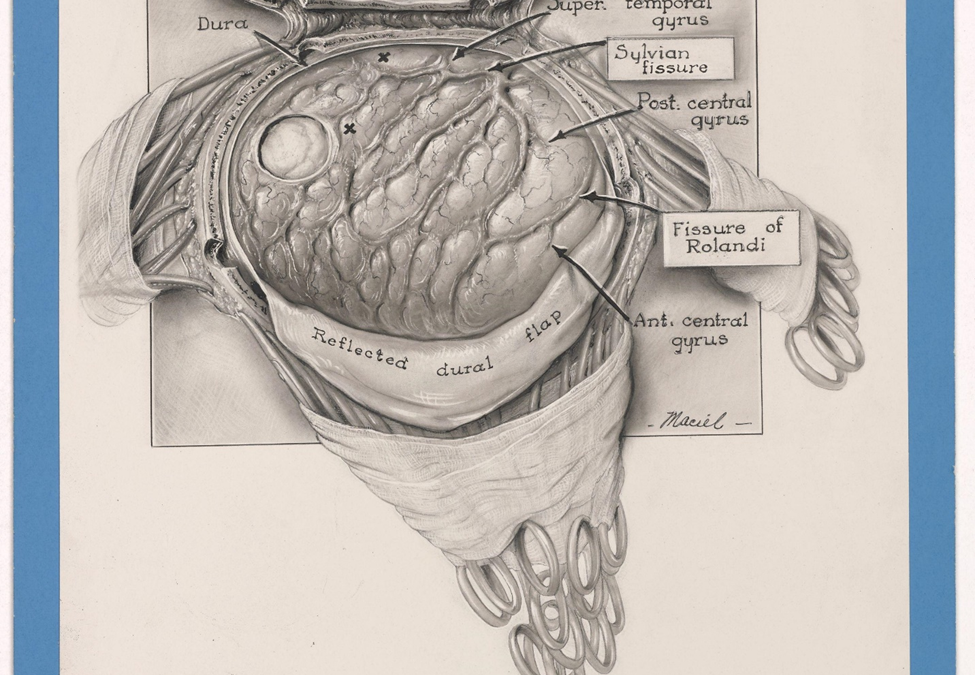
Brain Surgery
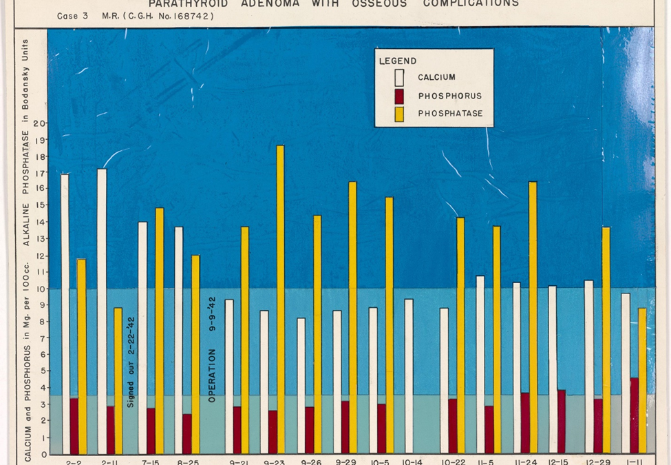
Parathyroid Adenoma Osseous
The Henry R. Winkler Center for the History of the Health Professions is grateful to James Van Mil, Sidney Gao and Sean Crowe for their expertise in the digitization of the Mary Maciel Archives.
ability to create personal accounts to access Board Review or NPTE review questions
Each collection also includes some unique features:
AccessPediatrics: calculators, algorithms, and a Clerkship Corner; textbooks, cases, and Q&A especially for medical students
AccessPhysiotherapy: topics by modalities and an interactive cadaver dissection experience in its Anatomy and Physiology Revealed section
AccessSurgery: images, videos, and descriptions of procedures.
Bookmark these new resources or go to the Health Sciences Library’s home page http://libraries.uc.edu/hsl/ and click on eBooks from the menu on the left under Quick Links or Point of Care eResources in the center of the page under Express Links.
If you have any questions, please contact Edith Starbuck at edith.starbuck@uc.edu or 558-1433.
We extended the AccessPediatrics, AccessPhysiotherapy, and AccessSurgery trials so more of you would have the chance to try out these resources and let us know what you think. Many thanks to those of you who have taken a look at the trials and filled out a survey.
Visit the HSL: Trials for New Electronic Resources guide to access the trials. Each trial includes a brief survey so you can add your voice to the decision-making process.
The electronic resources currently under consideration are the following: